Small Animal Internal Medicine
Radiomics & Ultrasound Imaging In Cats Can we Distinguish IBD vs. LSA without A Biopsy?
Radiomics & Ultrasound Imaging in Cats Can We Distinguish IBD vs. LSA Without a Biopsy?
Friday, June 16, 2023
1:30 PM - 2:30 PM ET
Location: PCC 113AB
CE: 1

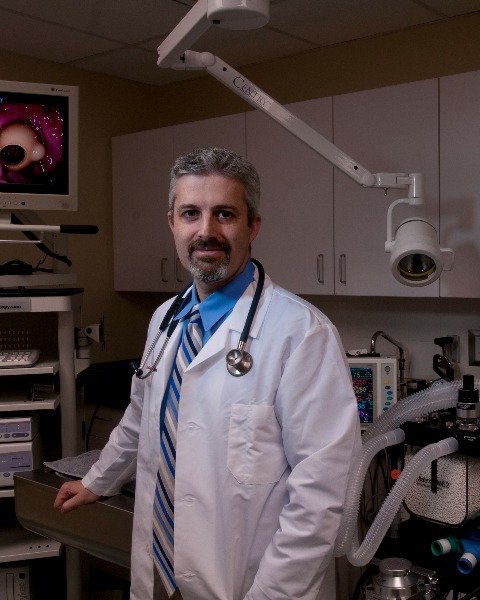
Marnin A. Forman, DVM, DACVIM (SAIM) (he/him/his)
Head of Internal Medicine
CUVS (Cornell University Veterinary Specialists)
Stamford, Connecticut, United States
Primary Presenter(s)
Presentation Description / Summary: Abdominal ultrasonography (US) is a non-invasive, rapid, accessible imaging modality commonly used as a diagnostic tool for abdominal diseases in cats. It is moderately sensitive in detecting intestinal disease but lacks specificity. It is operator/observer skill and equipment dependent. Manual, ultrasound-based measurements of intestinal total wall thicknesses, or ratio of the muscularis to the submucosa layers, can aid in the recognition of intestinal diseases but does not distinguish between inflammatory bowel disease (IBD), small cell epitheliotropic lymphoma (SCEL) and other GI disorders. Ultrasound images contain a large amount of data, and some of the data is difficult for the human eye to detect. Artificial Intelligence (AI) may be able to provide decision support in interpreting US images and ultimately reach a diagnosis without requiring histopathology. Quantitative image biomarkers, or radiomics, from US images, are used in the development of machine learning models. Initial work in cats, establishing "ground-truth" data for training AI systems, has been successful and reported. In ultrasound images from normal cats and cats with IBD or SCEL, segmentations (the perimeter of the intestines with a single closed region of interest) has lower intra- and interobserver repeatability and agreement than thickness measurements of the small intestine layer. This talk will discuss the use of intestinal ultrasound radiomics in cats and AI and the potential role of AI in Veterinary Medicine.
Learner Outcomes: 1. The use of artificial Intelligence has been studied in Human and Veterinary Medicine and may be utilized more frequently in the future and permit less invasive testing to reach a diagnosis. 2. Ultrasound images contain a large number of quantitative image biomarkers, termed Radiomics, that can be difficult for the human eye to detect and are used in the development of machine-learning models
Learner Outcomes: 1. The use of artificial Intelligence has been studied in Human and Veterinary Medicine and may be utilized more frequently in the future and permit less invasive testing to reach a diagnosis. 2. Ultrasound images contain a large number of quantitative image biomarkers, termed Radiomics, that can be difficult for the human eye to detect and are used in the development of machine-learning models
Learning Objectives:
- Upon completion, the participant will be able to appreciate the utilization of quantitative image biomarkers, or radiomics, in feline GI ultrasound images.
- Upon completion, the participant will be able to understand how clinical GI measurements (i.e., total wall thickness) correlate with radiomics.
- Upon completion, the participant will be to describe studies utilizing radiomics in cats diagnosed with IBD or small intestinal lymphoma and propose situations in which radiomics can be used in place of obtaining GI biopsies.

